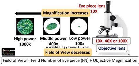
field of view for microscope|how to use a light microscope : iloilo The microscope field of view is found with the following formula: Field of View = Field Number (FN) ÷ Objective Magnification. If an auxiliary lens is being used on a stereo microscope, the magnification factor of this . Revolution energy saving design with Easy Energy Saver technology; Supports newest NEC SuperSpeed USB 3.0 with superfast transfer rates ; 3X USB power delivery for greater compatibility and extra power for USB devices

field of view for microscope,A microscope’s field of view (FOV) helps determine the approximate size of objects too small to measure with a ruler. To calculate the field of view diameter, divide the field number by the magnification .Field of View. The diameter of the field in an optical microscope is expressed by the field-of-view number, or simply the field number, which is the diameter of the view field in .To calculate a microscope’s field of view (FOV), it’s necessary to know its field number and objective magnification. Learn more about FOV and its importance.

The microscope field of view is found with the following formula: Field of View = Field Number (FN) ÷ Objective Magnification. If an auxiliary lens is being used on a stereo microscope, the magnification factor of this . In a microscope, the microscopy field of view is the diameter of the viewing field measured at an intermediate plane of angle. To put it simply, it’s the diameter of the .
Field of view (also abbreviated as FOV) for a microscope is the extent of the observable area in distance units. The optics provide a clear and undistorted view in a field around .
Field of view or FOV is the area of the object that is imaged by a microscope system. The size of the FOV is determined by the objective magnification. When using an eyepiece .

What is this “FIELD of VIEW”? In a technical sense, we are talking about the diameter of the INTERMEDIATE IMAGE, created by the objective or, in infinity systems, . The microscope field of view, or field diameter, is the distance across the image as seen through the microscope. The field of view is greater at lower magnifications. For example, at 10x .Field of View or Field Diameter is very important in microscopy as it is a more meaningful number than "magnification". Field diameter is simply the number of millimeters or micrometers you will see in your whole field of view when looking into the eyepiece lens. . Zoom microscopes have a fixed working distance throughout the zoom range. When .Field of View. Field of view or FOV is the area of the object that is imaged by a microscope system. The size of the FOV is determined by the objective magnification. When using an eyepiece-objective system, the FOV from the objective is magnified by the eyepiece for viewing. In a camera-objective system, that FOV is relayed onto a camera .Example 1: Calculate the field of view diameter of an optical microscope with a 45× objective lens, eyepiece field number 15 and without a tube lens (its magnification is 1×). Example 2: Calculate the field of view diameter for a 45× objective lens if the field of view for an objective lens 5× is 3 mm. Example 3: Estimate the size of a .
Microscope field of view (FOV) is the maximum area visible when looking through the microscope eyepiece (eyepiece FOV) or scientific camera (camera FOV), usually quoted as a diameter measurement (Figure 1). Maximizing FOV is desirable for many applications because the increased throughput results in more data collected which gives a better .The field of view is the maximum area visible through the lenses of a microscope, and it is represented by a diameter. To determine the diameter of your field of view, place a transparent metric ruler under the low power (LP) objective of a microscope. Focus the microscope on the scale of the ruler, and measure the diameter of the field of .The 40mm virtual field of view of the ASOM is compared to that offered by a traditional microscope using a 1024×1024 and 4096×4096 camera (all systems operating at 0.21 NA). The 0.38mm size of the ASOM sub-field of view is also shown with a 512× 512 camera, requiring many scan movements to cover the entire 40mm field. The microscope field of view calculator is a valuable asset for the scientific community. From its definition to its applications, it serves a wide range of purposes across different fields. It stands as an essential tool that facilitates precision, accuracy, and reliability in various scientific endeavors, thereby holding a significant place .
A fixed focal length lens, also known as a conventional or entocentric lens, is a lens with a fixed angular field of view (AFOV). By focusing the lens for different working distances (WDs), differently sized field of view (FOV) can be obtained, though the viewing angle is constant. AFOV is typically specified as the full angle (in degrees .
The limit of resolution of a standard brightfield light microscope, also called the resolving power, is ~0.2 µm, or 200 nm. Biologists typically use microscopes to view all types of cells, including plant cells, animal cells, protozoa, algae, fungi, and bacteria. With a 5X objective, the 1 mm scale fits 5 times within the diameter of my microscope, therefore the field of view is 5 mm or 5,000 microns. At 10X the scale fits 2.3 times so the field is 2.3 mm or 2300 microns. With a 20X objective the field of view is 1,150 microns and at 40X it’s 560 microns (on my microscope).
3. Determine the field of view: The field of view is the visible area when looking through the microscope. It is determined by the diameter of the field diaphragm and the magnification of the objective lens. To determine it, use a ruler or a micrometer to measure the diameter of the field diaphragm. 4.
how to use a light microscopeThe field number (F.N.) is referred to as the diaphragm size of eyepiece in mm unit which defines the image area of specimen. The diaphragm diameter actually seen through eyepiece is known as the practical field of view (F.O.V.) which is determined by the formula: > Top of Product page. > Top of Digital Microscope page.field of view for microscope how to use a light microscopeThe field of view (sometimes abbreviated FOV), which is visible and in focus when observing specimens in a microscope, is determined by the objective magnification and the size of the fixed field diaphragm in the eyepiece. When the magnification is increased in either a conventional or stereomicroscope, the field of view size is decreased if . The field of view (FOV) in microscopy is a critical parameter that determines the size of the observable area. It is calculated using the formula: Field of View = Field Number (FN) ÷ Objective Magnification. This guide provides a detailed explanation of the field of view formula, its theoretical background, measurable data, figures, data .field of view for microscopeThe field diameter in an optical microscope is expressed by the field-of-view number or simply field number, which is the diameter of the viewfield expressed in millimeters and measured at the intermediate image plane.The field diameter in the object (specimen) plane becomes the field number divided by the magnification of the objective. There is an inverse relationship between magnification and diameter of field. Place a thin, clear, metric ruler on the stage. Hold it in place with the stage clip. Use the scan objective to focus and observe the millimeter marks on the ruler. Record the diameter of field using scan objective lens in mm. Include half spaces. Convert to micrometers.The field diameter in an optical microscope is expressed by the field-of-view number or simply field number, which is the diameter of the viewfield expressed in millimeters and measured at the intermediate image plane. The field diameter in the object (specimen) plane becomes the field number divided by the magnification of the objective. Introduction to Widefield Microscopy. One of the basic microscopy techniques is known as ”widefield microscopy”. Fundamentally, widefield means the entire specimen or sample is exposed to the light source with the resulting image being viewed by the observer either via the eyepieces or on a monitor connected to a digital camera.
field of view for microscope|how to use a light microscope
PH0 · how to use a light microscope
PH1 · history of microscopy
PH2 · different types of microscopes
PH3 · Iba pa